 ALERT
ALERT ATTENTION ⚠️️
Maintenance has been scheduled for iLab Primary U.S. on June 13, 2026 at 3:30PM (UTC). During this time, the iLab platform will be inaccessible or limited in functionality. Details and updates can be found on the Agilent Status Page.
For additional information, please contact the Agilent iLab Support at ilabsupport@agilent.com.
https://digislide.app.vumc.org
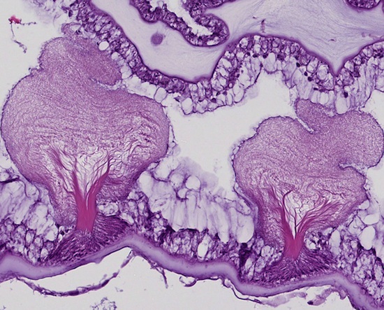
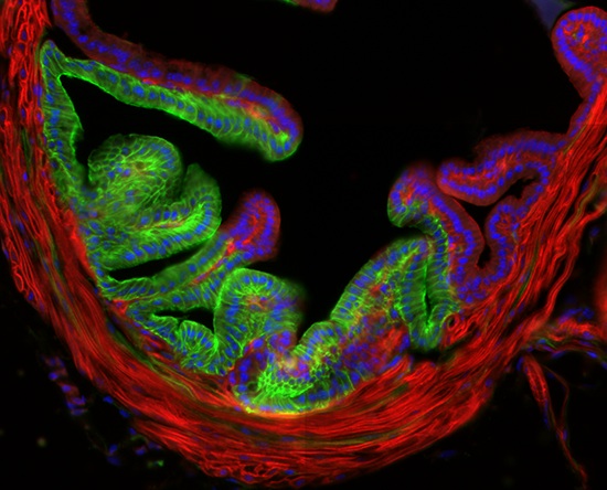
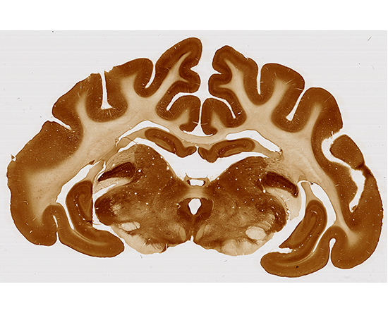
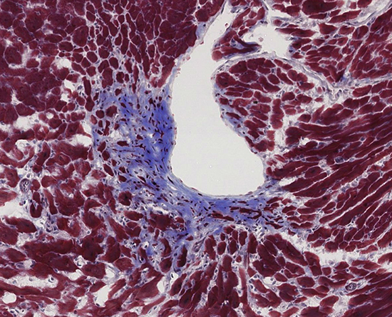
The Digital Histology Shared Resource provides large-scale digital archiving and quantitative analysis of histologic, immunohistochemical and immunofluorescence staining of tissue sections and tissue microarrays and houses an array of sofisticated whole slide imaging platforms:
Aperio Versa 200 (Leica/Aperio) Automated Slide Imaging System for bright field and fluorescence whole-slide imaging. This platform holds up to 200 slides per batch; has 6 separate filter channels: DAPI, Cy2, Cy3, TxRed, Cy5, and Cy7. It is the ideal system for rapid, automated, and consistent whole-slide imaging in a wide range of fluorescent channels and bright field modalities. The DHSR operates two identical Aperio VERSA 200 systems, alowing for double the throughput.
eSlideManager (Leica/Aperio) web-based digital whole slide review, sharing, annotation, and analysis platform. This software is the modern upgrade to the aging Digital Image Hub with tighter cybersecurity, better searching and desktop features. This software is capable of auto uploading images directly from the Aperio AT2 400 into its database.
Analysis and computational power: The DHSR hosts ~150TB of whole slide images on the VUMC petabyte array, as well as over 150TB of local RAID6 storage to gaurd against data loss. Additionally, five HP workstations (Z8-series) with large format screens (~50”) are utilized by the DHSR for image quantification and export. The automated imaging and analysis performed in this core saves researchers and staff weeks of tedious work. An additional service offered by the DHSR is the creation of digital archives of critical and irreplaceable tissue samples, a benefit only feasible due to the automated high-resolution imaging of whole 25 mm x 75 mm microscope slides and 50 mm x 75 mm slides.
GelCount system by Oxford Optronix scans and counts mammalian organoids and yeast or bacterial colonies in a wide variety of Petri dish and cell culture plate formats. This system is designed for the detection, counting and characterization of stained/adherent mammalian cell colonies or of unstained/non-adherent colonies in soft agar or collagen assays, but also works very well for yeast and bacterial colonies. The software is fully trainable and can be programmed to recognize specific colony features. Detailed information such as diameter, area, density, and nearest neighbor is provided, as well as high-resolution images.
To Submit Slide Submission Requests: Click the Request Services Tab
To Reserve an Instrument: Click the Schedule Equipment Tab
DHSR Website: https://www.vumc.org/dhsr/
Digital Slide Archive: https://digislide.app.vumc.org
eSlideManager: https://eslidemanager.app.vumc.org
Scientific Director: James R. Goldenring, M.D., Ph.D.
Managing Director: Joseph T. Roland, Ph.D.
| Hours | Location |
|
Monday-Friday |
10425 MRB IV |
For whole slide imaging and quantification:
Whole slide imaging and quantification of immunostaining were performed in the Digital Histology Shared Resource at Vanderbilt University Medical Center (www.mc.vanderbilt.edu/dhsr).
For whole slide imaging:
Whole slide imaging was performed in the Digital Histology Shared Resource at Vanderbilt University Medical Center (www.mc.vanderbilt.edu/dhsr).
For colony counting:
Colony imaging and quantification was performed on the GelCount System (Oxford Optronix) in the Digital Histology Shared Resource at Vanderbilt University Medical Center (www.mc.vanderbilt.edu/dhsr).
| Request Services or View Current Rates |
| ► Leica Brightfield (1) | |||
| Name | Description | Price | |
|---|---|---|---|
| 20X Brightfield Scanning |
Charged per slide |
Inquire | |